Microscopy
60 readers
1 users here now
Community for identifying and discussing microphotographs as well as microscopy techniques, news and microbial processes in general.
Microscopy is the technical field of using microscopes to view subjects too small to be seen with the naked eye.
There are three well-known branches of microscopy: optical, electron, and scanning probe microscopy, along with the emerging field of X-ray microscopy.
Researcher or microscope owner with some cool pics? Share it! Saw a weird critter in your pond samples the other day? Share it!
🍃🌻 Rule 1: Kindly be empathetic and kind to others. Trolling and spamming will not be tolerated. Making the community a friendly & supportive place is our goal.
All of us are here for a short lifetime. Let’s have a nice time here and avoid negativity. :)
founded 2 months ago
MODERATORS
2
5
6
7
10
11
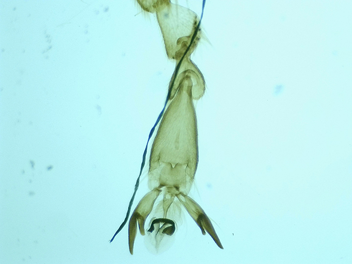
Taken with my SW350T Compound Trinocular Microscope. Taken at 10x. Domestic honeybee (Apis mellifera) leg and claw.
12
Taken with my SW350T Compound Trinocular Microscope. Taken at 10x. Hornwort (Ceratophyllum demersum) stem
